Patient Education
Treatments

Trigger Point Injections
Used for muscle tightness and muscle injury in conjunction with a stretching program. Involves the injection of local anesthetic (and/or steroid/Botox) when appropriate.

Physical Therapy
Used to improve mobilization, strengthening and range of motion, mobilization, stabilization and strengthening. Ultrasound, electrical stimulation, ice, heat, bracing and exercise are utilized by trained physical therapists.
Epidural Steroid Injections
Injections of local anesthetic and steroid to target an inflamed spinal nerve or disc under fluoroscopic (x-ray) guidance. Often used for sciatica, pinched nerves, radiculopathy, degenerative disc disease, bulging or herniated discs. Can also be referred to as a nerve block or selective root block. May aid in diagnostic workup or therapeutic treatment. Can be accomplished in the cervical (neck), thoracic (mid back) and the low back (lumbar) segments. May be accomplished with sedation when requested.
Facet Blocks / Facet Injections
Targeting of the joints and their innervations to help diagnose pain generation from spinal joints in the cervical (neck), thoracic (mid-back), lumbar (low back) segments. This is commonly referred to as mechanical spine pain. Some forms of sciatica and radiculopathy are actually a referred pain from a spinal joint that can mimic a pinched nerve. Accomplished under fluoroscopy (x-ray) and sedation when requested.
Radiofrequency Lesioning / Rhizotomy / Ablation
Used after a diagnosis of joint pain generators are made for more long-lasting results. Small lesions using radio waves are applied by small needles to destroy pain fibers in the joints of the spine (facets), knees and hips. Fluoroscopic (x-ray) guidance is used with sedation when requested.
Neuroplasty / Lysis of Adhesions
Catheter guided placement of epidural steroids and enzymes to break up surgical and non-surgical adhesions in the epidural space. This allows greater mobility of an impinged nerve root by adhesive material. Used for post laminectomy syndrome, sciatica, radicular pain, and degenerative disc disease. Can be used in the cervical (neck), thoracic (mid-back), lumbar(low back) segments. Utilizes fluoroscopy (x-ray) and sedation when requested.
Sacroiliac Joint Injections
A concentrated dose of local anesthetic and steroid to reduce inflammation within the joint is used for diagnosis and treatment with the aid of fluoroscopy (x-ray).
Stellate Ganglion Blocks
Are you suffering from Parosmia, Anosmia, Dysgeusia, or long COVID symptoms? We can help. We perform stellate ganglion blocks under fluoroscopic guidance for treatment of this debilitating condition.
Dr. Andrew Goldberg and Dr. Seth Wachsman are board certified in both Pain Management and Anesthesiology. They have been performing stellate ganglion blocks for more than 25 years each.
Stellate Ganglion Blocks are injections for sympathetic pain issues in the upper extremity (complex regional pain syndrome (CRPS) and reflex sympathetic dystrophy (RSD). The blocks assists with diagnostics and therapeutic treatment. These injections are placed under the skin in the anterior neck with x-ray (fluoroscopy) and with sedation, identify and treat sympathetic symptoms after trauma and surgery as well as peripheral vasculature disease and Raynaud’s Syndrome including pain, swelling, color change, temperature change, hair and nail bed changes and inability to touch the effected limb.
Lumbar Discogram / Cervical Discogram
The placement of a needle into the center of a disc to determine whether the disc is a possible pain generator. Procedure is accomplished with x-ray (fluoroscopy) and sedation. Contrast is placed in center of the disc (nucleus) to assess for leaks and pain reproduction as well as measuring the pressures in the disc. Allows for further surgical and non-surgical treatment planning. Used for disc herniation, disc bulge, degenerative disc disease.
Spinal Cord Stimulator
Placement of electrodes through a needle to block pain signals from the spine and extremities. Used in the lumber (low back), thoracic (mid back) and cervical neck segments.
A 7-day trial begins with a small electrode (wire) placed with an epidural needle and while the needle is removed the electrode is left in the epidural space and attached to an external power source. The initial electrode is removed easily after 7 days in all cases. If the trial is deemed successful, then a new set of electrodes are place with or without a generator (pacemaker battery) or a Bluetooth setup for a ore permanent placement.
The electrodes are controlled by a small handheld controller that the patient can carry. Used for post-laminectomy syndrome in all areas of the spine, failed back syndrome, complex regional pain syndrome/ reflex sympathetic dystrophy, peripheral neuralgia, extremity nerve injury and pain from peripheral vasculature.
Lumbar Sympathetic Blocks
Injections on the side of the lumbar spine to assist diagnosis of sympathetic pain issues (complex regional pain syndrome (CRPS) and reflex sympathetic dystrophy (RSD). These injections help lower extremity pain that presents with limb pain, color change, temperature change, swelling, inability to touch the limb, loss of hair and nail bed changes.
They can additionally assist therapeutically and be used in combination with radio frequency lesioning if necessary for longer acting relief. Can occur after surgery or trauma to the limb. Accomplished under x-ray (fluoroscopy) and with sedation. Can additionally be used for peripheral vasculature disease pain.
Peripheral Nerve Stimulator
For pain relief to the shoulder, arm, wrist, hand, leg, and knee when most other procedures have failed. Surgery may have already been performed. This approach allows us to place a specialized catheter under the skin and adjacent to the irritated or abnormal nerves. The device sends a painless electric impulse across several layers of tissue until it reaches the painful nerves. The electric impulse suppresses the painful nerves and can minimize or eliminate the pain.
This is a two step process. The first part includes a temporary trial, lasting about five days. The second part includes permanent placement of the catheter, with the excess placed under the skin. With the approach used, this type of treatment is always reversible, with removal of the catheter/lead if necessary.
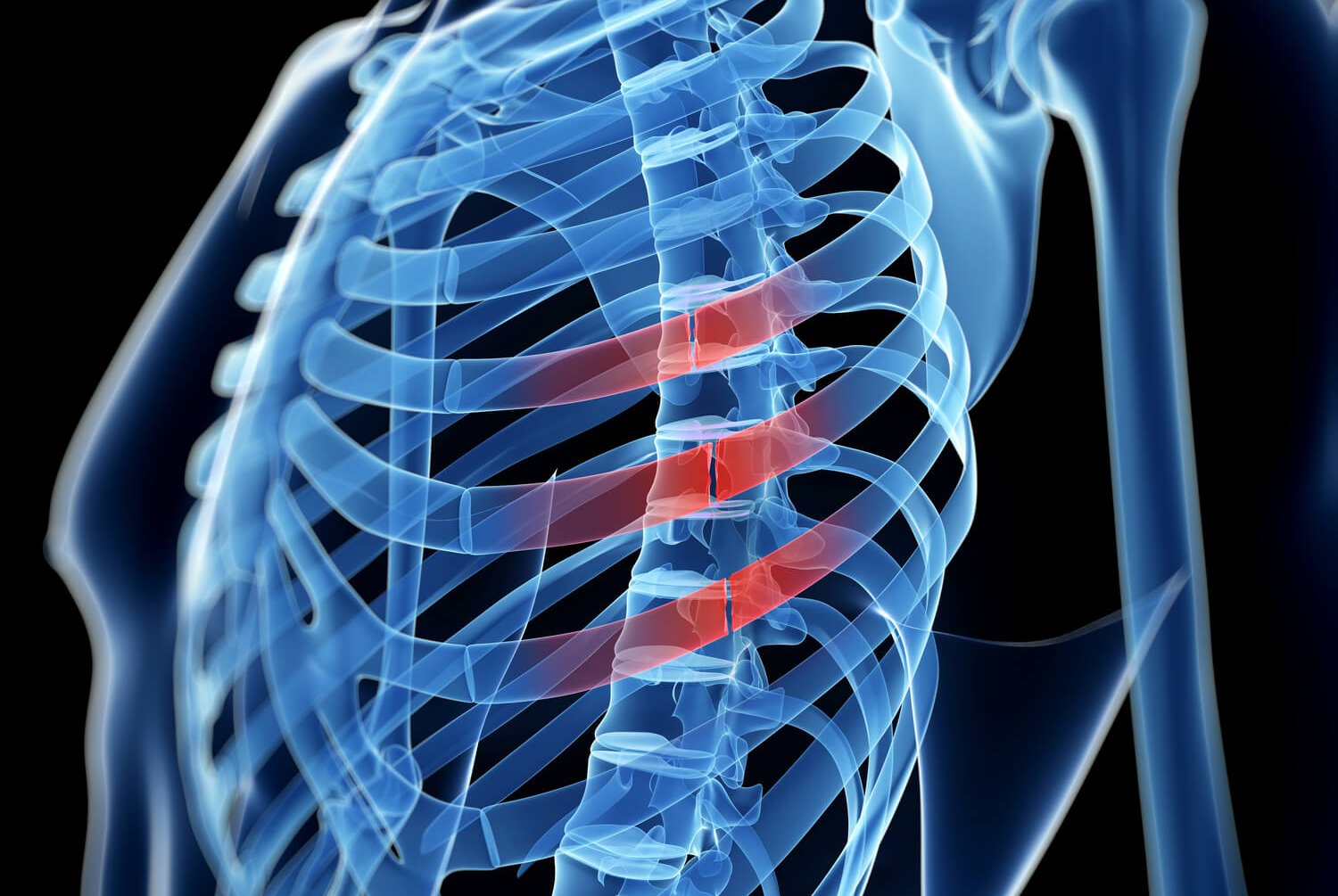
Intercostal Blocks
Used for chest wall pain from trauma/rib fracture. Used for diagnostic to distinguish from abdominal issues versus abdominal wall issues. Utilizes fluoroscopy (x-ray) and sedation when requested.
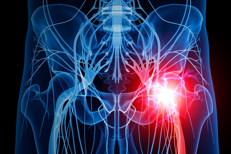
Peripheral Nerve Blocks
Nerves such as the ilioingual (groin pain/post hernia surgery), lateral femoral cutaneous nerve (thigh pain), sciatic/piriformis (leg pain), suprascapular block (shoulder). Fluoroscopic (x-ray) guidance utilized along with sedation.
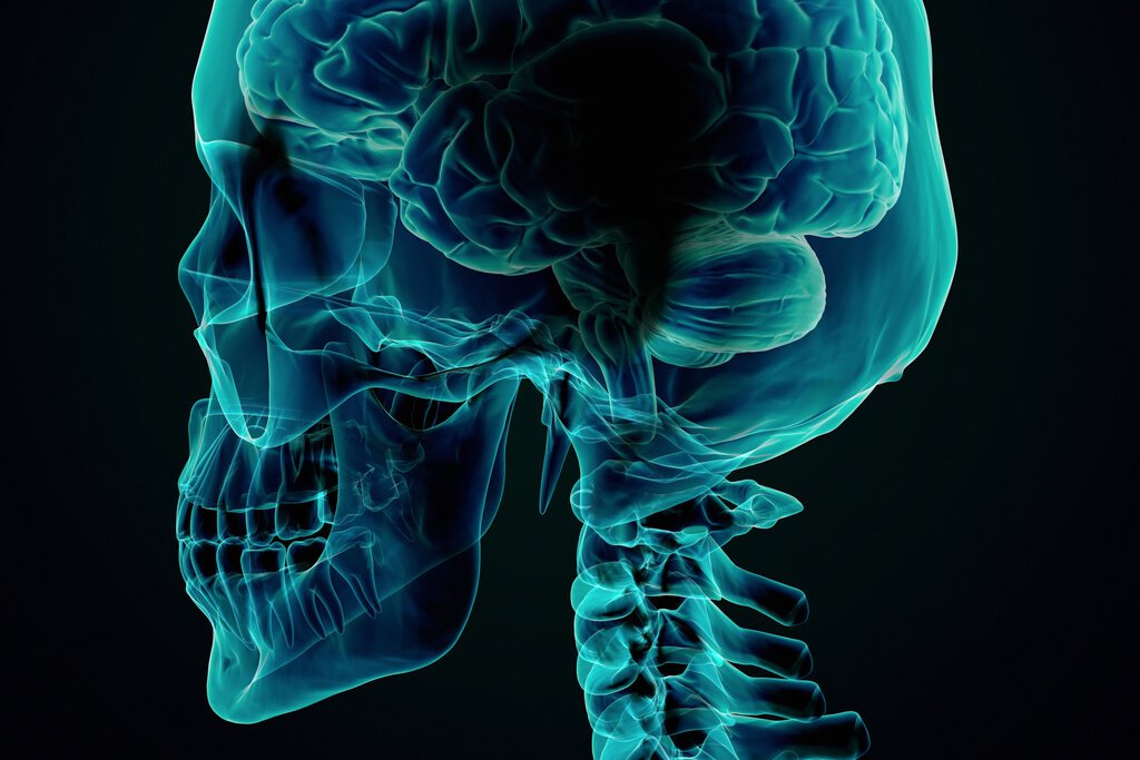
Cranial Nerve Blocks
Nerve blocks used for trigeminal neuralgia and facial pain. Fluoroscopic (x-ray) guidance utilized along with sedation.
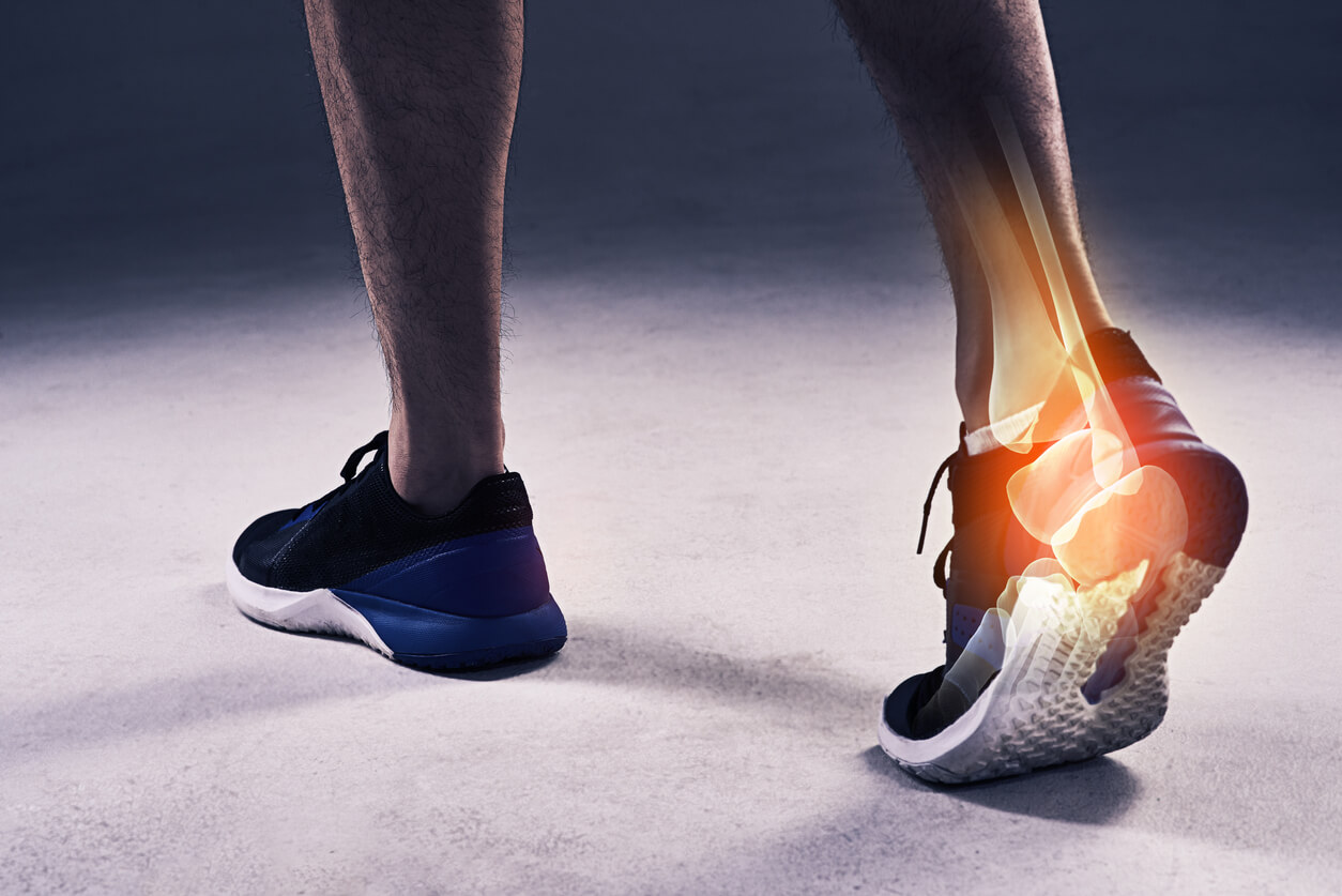
Joint Blocks / Arthrography
Injections in the shoulder, hip, knees, wrists, ankles for diagnostic and therapeutic purposes. Can utilize fluoroscopy or ultrasound guidance.
Center for Medical Marijuana
The advantage of using medical marijuana over narcotics is providing pain relief while AVOIDING the unpleasant side effects of constipation, nausea, and respiratory depression. You are unlikely to experience any “high” from the use of medical marijuana.
Learn More

